Dental X-Rays in Turkey

What are dental X-Rays and why are they needed?

X-rays show the position of the teeth, roots, condition of the jaw and facial bones, helping dentists find the problem. X-rays pass through or be absorbed solid bodies at different intensities, allowing different tissues to appear and be distinguished from the dental film in different colors. It helps to find and solve problems that cannot be found by examination. It also gives information about the success of the treatments performed.

What types of problems do X-Rays help detect?

X-rays are used to diagnose problems in the teeth and jaws.

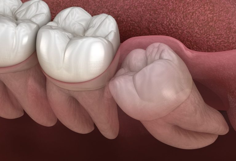
Especially small areas of caries between the teeth and decay under the existing fillings, bone loss in the jaws, the proximity of the area to be treated to anatomical structures, abscesses, cysts, tumors, suitability of dental implants, impacted teeth

What are the types of dental X-Rays in Turkey?

There are two types of dental X-Rays: intraoral(the X-Ray film is inside the mouth) and extraoral (the X-Ray film is outside the mouth)

Dental Treatment with General Anesthesia Sedation in Turkey

The most common type of X-ray; Intraoral X-Rays shows different aspects of the teeth.

Bitewing X-rays show details of the upper and lower teeth from their crown in one area of the mouth. Thanks to this, decay between the teeth and gum diseases are detected. It also shows the proper fit of crowns and bridges with the gum and the wear of fillings.

Periapical X-Rays show the entire tooth and root. It detects changes in the root and surrounding bone structure.

Occlusal X-Rays show the entire arch of the teeth in either the upper or lower jaw.

Are dental X-Rays safe in Turkey?

The amount of radiation emitted from X-rays is low. The amount of radiation emitted by technological developments such as high-speed X-rays, the use of lead-lined aprons is reduced. Despite the safety of X-Rays, no X-Rayed if it is unnecessary.

Extraoral X-Rays are used to detect dental problems in the jaw and skull.

Panoramic X-Rays show the entire mouth area; all the teeth in the upper and lower jaws, sinuses, jaw joint connecting the lower jaw to the skull.

Cephalometric projections show an entire side of the head. This X-ray allows to examine the appearance of patients from the jaw and profile. It is often used by orthodontists.

The sialogram shows the diagnosis of diseases such as Sjogren’s syndrome, which may be in the salivary glands, the causes of dry mouth and soft tissues that may not be seen on the X- ray.

CT and CBCT are types of 3D imaging. It is used in the placement of dental implants, to evaluate cysts and tumors in the mouth and face. 3D type of dental imaging techniques provides more reliable information and high-quality images than the 2D type of dental imaging techniques.

MRI that takes a 3-D view of the oral cavity imaging is ideal for soft tissue evaluation.